I love a science headline that makes me question whether I arrived with all the parts listed in the instruction manual. Scientists finding a possible new organ is especially good for that. You'd think we'd have a fairly complete inventory of what we're carrying around by now, considering how much effort we've put into examining ourselves. Apparently, there's still room for a surprise between the organs we already knew about.
The interstitium caught my attention for a slightly different reason, though. Part of the story involves the way tissue changes when we prepare it to be studied. A structure can look different on a slide from the way it looks while it's still full of fluid, and that difference helped send researchers back to something familiar. I find that wonderfully inconvenient. Even the act of getting a better look can alter the thing we're trying to understand.
First, the fluid around our cells
The word interstitium refers to spaces and supporting material between structures in our tissues. Interstitial fluid occupies the spaces around cells, where it helps deliver oxygen and nutrients and carry away waste products. The National Cancer Institute's definition connects that fluid with material moving out of the smallest blood vessels, the capillaries. Once interstitial fluid enters lymphatic vessels, we call it lymph. The name changes with where the fluid is.
There are several fluid compartments to keep track of. Fluid inside cells is intracellular, while fluid outside them is extracellular; blood plasma and interstitial fluid are different parts of that outside compartment. OpenStax's anatomy text describes water and dissolved substances moving among these spaces. So the underlying idea of fluid around cells was already part of physiology. The surprising question concerned the architecture of larger spaces within tissues.
Connective tissue has quite a lot happening in it
I think connective tissue has been given a rather unfortunate name. It sounds like the packing material that arrives around something more exciting, even though it's doing essential work. As OpenStax explains, its cells sit within an extracellular matrix, material outside the cells that includes a ground substance and protein fibers. Different arrangements give different connective tissues their properties. The tissue helping support an organ and the tissue forming a tendon have different jobs to do.
That combination of fluid, fibers and cells matters to the story. Describing something as a space can make it sound empty, like the gap between two books on a shelf. Here, the surroundings are part of how the tissue works. I also like the change in perspective involved: instead of looking only for an interesting cell, we're looking at the material around it and asking what that setting allows to happen.
What the researchers saw in 2018
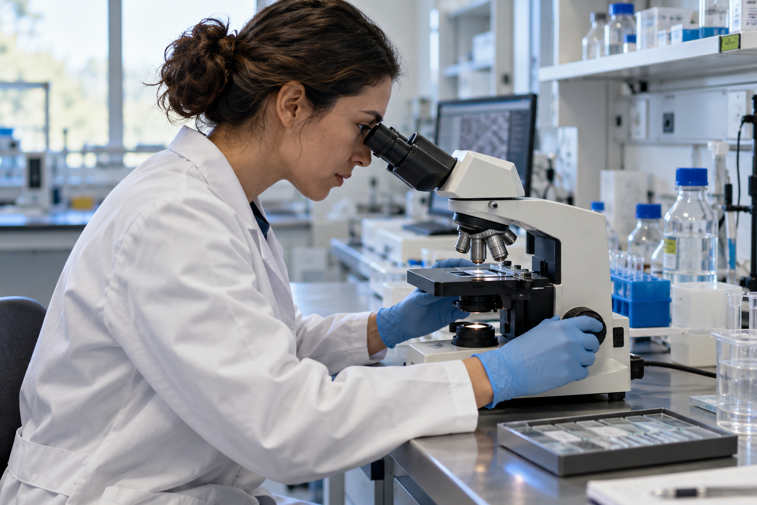
In a 2018 paper in Scientific Reports, Petros Benias, Rebecca Wells and colleagues described an unusual pattern seen while examining bile-duct tissue with probe-based confocal laser endomicroscopy. The technique lets researchers obtain microscopic images during an endoscopic examination. After fluorescent dye was given, they saw bright, fluid-filled spaces separated by dark branching bands. The pattern needed an anatomical explanation.
Comparing fresh-frozen tissue with routinely processed specimens helped supply one. The team found spaces supported by thick collagen bundles; ordinary processing could let fluid escape and leave the arrangement collapsed. Similar structures appeared in other tissues, including skin, fascia and parts of the digestive tract. The paper expanded the description of the interstitium rather than introducing the general idea that tissues contain fluid.
This is the detail that makes the discovery memorable for me. We tend to treat a scientific image as a straightforward picture of what's there, but producing the image involves decisions about preparation and preservation. A slide can tell us an enormous amount while still leaving another feature harder to recognize. I find that considerably more interesting than the idea that everybody somehow forgot to look under the skin for several centuries.
How the “new organ” description entered the story
Pathologist Neil Theise, one of the study's senior authors, has argued that the widespread network deserves to be considered an organ. In a 2018 NYU Langone interview, he pointed to its recurring structure and proposed functions as reasons for that view. He also described organ status as something that develops through the scientific community's treatment of a structure over time. That is a classification argument alongside the anatomical observations.
Theise proposed that compressible, fluid-filled spaces could help absorb mechanical forces as tissues move. Consider how often the digestive tract changes shape while doing its job, or how repeatedly the lungs expand. A possible cushioning role is a useful physical hypothesis to investigate. It needs evidence about forces and tissue behavior, rather than the much broader claim that a previously hidden organ explains every physical impact or emotional hurt.
I understand why the organ label attracted so much attention. Finding another item for the anatomy list sounds wonderfully dramatic, and it gives the story a very tidy hook. My interest is in what the spaces do, how they're connected and what we can measure within them. Those questions remain worthwhile whichever label eventually becomes most useful. A discussion over the filing system doesn't make the contents of the folder dull.
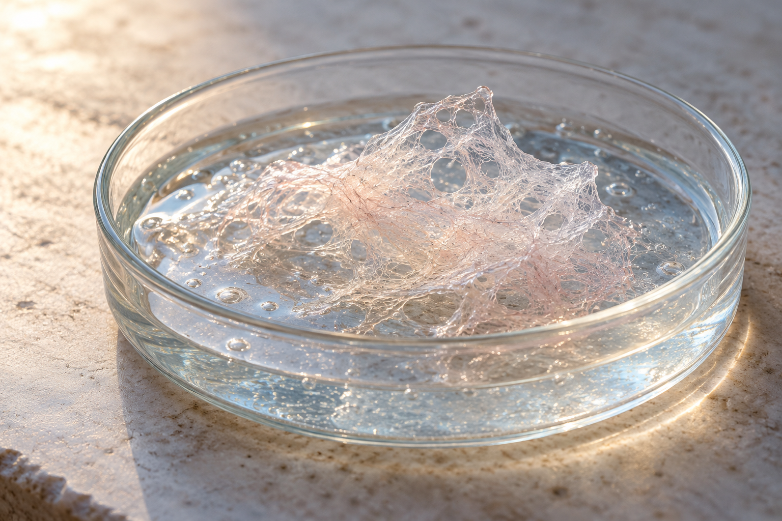
Editorial concept showing a fiber mesh in gel. This is a material analogy, rather than a microscopic image of tissue.
Tattoo pigment became a clue to the connections
A 2021 follow-up study investigated whether interstitial spaces continued across tissue and organ boundaries. Odise Cenaj and colleagues examined the locations of particles, including tattoo pigment, and used staining to identify hyaluronic acid, a component of these spaces. Their observations in tissues such as skin and colon supported connections extending into adjacent connective tissue and along the coverings of nerves and blood vessels. Tattoo pigment is a wonderfully unexpected thing to find in an anatomy investigation.
The study also raised questions about how these routes might participate in the movement of cells and other material, including the spread of cancer. Those are implications to investigate, rather than proof of one universal route for every tumor. Continuity between spaces tells researchers something about possible paths. Working out which cells use them, under what conditions and with what consequences adds another set of questions.
My curiosty tends to wander toward those practical details. A map showing that two places connect is interesting, but I'd immediately want to know what travels between them, how quickly it gets there and what changes along the way. In this case, those are questions people can approach with tracers, stains and samples. There's something satisfying about a large, rather abstract claim becoming a series of small things somebody can actually look for.
Where the lymphatic system fits
The connection with lymph deserves a little room because it's easy to bundle all these spaces together. The lymphatic system collects fluid from tissues and returns it to the bloodstream, while lymph nodes provide sites for immune activity. OpenStax's account of the system describes small lymphatic vessels taking up interstitial fluid, then joining larger vessels. Fluid around cells and fluid inside a lymphatic vessel occupy different anatomical settings, even though they're connected by that movement.
That drainage matters when the system is disrupted. Fluid can accumulate in tissue spaces and produce swelling, including when lymphatic routes are damaged or obstructed. It's a real physical problem involving fluid balance and transport. Describing all of it as stagnant emotion loses the very features that make it possible to study, such as where fluid collects and which pathways are affected.
The lung study makes the larger question more specific
In a 2025 study of lung tissue, researchers examined specimens from nine patients and compared their findings with mouse lung tissue. Staining for hyaluronic acid and other markers showed continuity among major lung compartments and connections along structures surrounding nerves and blood vessels. This added evidence about how spaces inside the lung link with spaces outside it. The proposed network was being investigated one organ and one anatomical boundary at a time.
The authors discussed possible implications for inflammatory cells, disease spread and signaling. Those suggestions go beyond the staining observations themselves, which is where the next experiments come in. I like that progression: a broad idea about connected spaces leads to a much more particular examination of the lung. It makes the story feel ongoing, with a great deal still to work out between the first striking image and an explanation of what every connection does.
Researchers are also collecting the fluid itself
A different 2025 study developed ways to collect interstitial fluid from intestinal tissue. Ellen Avery and colleagues first worked with rodents, then adapted a tissue-elution method for use with samples from people. They measured substances including proteins, immune signaling molecules and metabolites produced by microbes. The amounts could differ between intestinal regions and from what was measured in blood plasma, giving researchers a more local view of the tissue environment.
That distinction interests me because a blood sample tells us something different from a sample taken close to where particular cells are interacting. The study presented a research approach, rather than a ready-made home test or a treatment. But it shows a concrete direction for the field: collecting material, measuring its contents and comparing places or conditions. Fluid that can sound like background scenery becomes something worth sampling carefully.
The current picture has several scales
In a 2026 review, Wells and Theise described interstitial spaces at several scales, from spaces between cells and around capillaries to larger spaces associated with fascia and other supporting tissues. Their focus included the surrounding matrix, fluid movement and signaling. The review also laid out open questions about what drives flow and how these spaces participate in communication. Even researchers advancing the broader network concept still have many details to resolve.
The word network is useful here, provided we keep asking what connects and how. A continuous structure can have local differences, and a route that exists anatomically can still have constraints on movement. I think the interesting work sits in those details. An entire-body connection is an exciting phrase, but finding out how the different parts behave is what gives the phrase substance.
Cells can respond to mechanical history
There is a real research term called mechanical memory, which is worth separating from memories of events in a person's life. In a 2014 experiment, researchers grew mesenchymal stem cells on surfaces with different stiffnesses. Previous time on a stiff surface influenced later cell responses, including responses after a change to a softer material. This was cellular behavior under controlled culture conditions. It wasn't evidence that connective tissue stores an argument, a bereavement or a childhood experience that can be released by pressing on it.
The distinction matters because words such as memory and signaling can acquire extra meanings as they move from a paper into an everyday conversation. A cell responding to its physical history is already fascinating. We don't need to give it a diary as well. The interstitium studies described here give us anatomy and possible mechanisms to investigate; they don't identify a hidden storage site for emotional trauma.
Questions about touch and acupuncture need their own evidence
Connective tissue research also attracts questions about massage, acupuncture and other practices involving touch or pressure. Those questions call for studies of the particular intervention and outcome. NCCIH's explanation of acupuncture describes several possible contributions, including effects involving the nervous system, local tissues and the context of treatment. Its mechanism is still being investigated. A newly described tissue arrangement doesn't by itself demonstrate that energy meridians have been mapped or that emotional experiences drain through interstitial fluid.
I understand the appeal of an anatomical explanation for something that feels physical. A feeling can be powerful without giving us a complete account of what caused it, though. Keeping the experience and the proposed mechanism distinct gives both a fairer hearing. It leaves room for someone's description of relief while also asking what an experiment actually measured.
The part that stays with me is how we look
I keep returning to the change in view that started this story. A familiar tissue looked different when researchers examined it in another condition, and that difference opened up questions about spaces, movement and connections. There's enough there to keep my attention without making the interstitium responsible for every mysterious sensation. The fluid, fibers and cells already give us a rather crowded set of things to understand.
I'm also fond of the idea that a place between the things we usually name can turn out to deserve its own careful examination. It's easy to be impressed by a heart, a lung or a brain and spend less time wondering about their surroundings. Now I'm interested in the surroundings too, including the unglamorous business of preserving a sample well enough to see them. Apparently, learning what's inside us still involves learning how to look.
Sources and further reading
Benias et al. (2018), structure and distribution of interstitial spaces
NYU Langone (2018), interview with Neil Theise on organ status
Cenaj et al. (2021), evidence for continuity across tissue boundaries
Ordner et al. (2025), interstitial continuity in and outside the lung
Avery et al. (2025), collecting intestinal interstitial fluid
Yang et al. (2014), mechanical memory in cultured stem cells